Loyola Core Microscopy Facility
Services Provided:
- Consultation
- Training
- Sample preparation
- Sectioning and staining
- Image acquisition
- Image analysis
Instrumentation
- Zeiss Elyra 7 dual iterative lattice structured illumination microscopy (SIM2) system
- Philips CM 120 transmission electron microscope
- Leica TCS SP5 multiphoton/confocal microscope
- Image analysis workstation
- Leica EM UC7 ultramicrotome
Contacts
Edward Campbell, Ph.D.
Director
708-216-3345
ecampbell@luc.edu
David Rademacher, Ph.D.
Manager
708-216-3395
drademacher@luc.edu
Loyola Core Microscopy Facility
Center for Translational Research and Education (CTRE)
Building 115, Rooms 173-176
2160 S. First Avenue
Maywood, IL 60153
Services
- Consultation
- Consult with principal investigators and graduate students to design experiments
- Share existing protocols
- Develop new protocols
- Training
- Training on all equipment
- Zeiss Elyra 7 dual iterative lattice structured illumination microscopy (SIM2) system
- Leica TCS SP5 multiphoton/confocal microscope
- Philips CM 120 transmission electron microscope
- Leica EM UC7 ultramicrotome
- Training of users on the preparation of samples for imaging by dual iterative lattice structured illumination microscopy (SIM2).
- Training of users on the preparation of samples for imaging by confocal microscopy.
- Training of users on the preparation of samples for imaging by transmission electron microscopy.
- Training on all equipment
- Sample Preparation
- Preparation of cell culture and tissue specimens for transmission electron microscopy. This includes fixation, osmification, dehydration, en bloc heavy metal staining, and embedding in epoxy resin.
- Preparation of virus, pseudovirus, protein, phage, extracellular vesicles, and synthetic nanoparticles for transmission electron microscopy. This includes incubating coated electron microscopy grids with the sample in solution then contrasting for subsequent imaging.
- Preparation of specimens, including tissue, protein, and extracellular vesicles, for immunogold electron microscopy.
- Preparation of specimens for correlative light and electron microscopy (CLEM) experiments.
- Preparation of specimens by photoconversion microscopy for imaging by transmission electron microscopy.
- Sectioning and Staining
- Sectioning of thin (~1 µm) and ultrathin (25-90 nm) sections via a Leica EM UC 7 ultramicrotome. This includes conventional ultramicrotomy and serial sectioning.
- Collection of thin sections, staining with toluidine blue, and imaging by light microscopy.
- Collection of ultrathin sections, including serial sections, onto electron microscopy grids.
- Staining samples with contrasting agents including lead citrate, uranyl acetate, and phosphotungstic acid.
- Performance of standard histological stains.
- Imaging Acquisition
- Multi-color imaging of live cells and fixed specimens.
- Imaging specimens by transmission electron microscopy.
- Single molecule localization microscopy (SMLM).
- Image Analysis
- Analysis of fluorescence by Zeiss Zen software, Leica Application Suite software, Oxford Instruments Imaris software, and Image J.
- Analysis of 3D volumes of electron micrographs via ESPINA software.
Pricing
- $30/hour for use of the Zeiss Elyra 7 dual iterative lattice structured illumination microscopy (SIM2) system.
- $30/hour for use of the Philips CM 120 transmission electron microscope.
- $25/hour for the use of the Leica TCS SP5 multi-photon/confocal microscope.
- $40/hour for sample preparation and staining.
- $20/hour for ultramicrotomy.
- No charge for consultation.
- No additional training charges. The cost of assisted and unassisted use of the instruments is the same.
- No charge for the use of the image analysis workstation.
- No charge for image analysis software.
- Services are to be paid with an account number through Portal or via invoice.
- Time on the instruments is to be reserved via Booked Scheduler https://ctre.bookedscheduler.com/Web/dashboard.php
Instrumentation

Dual Iterative Lattice Structured Illumination Microscopy (SIM2) with the Zeiss Elyra 7
Location
Center for Translational Research and Education (CTRE), room 175
Capabilities
- The Elyra 7 Lattice SIM2 system is versatile, allows for superior temporal and spatial resolution, is more efficient, and has a larger field of view than other systems.
- The Elyra 7 Lattice SIM2 system allows users to achieve resolutions of 60 nm (X, Y) and 200 nm (Z) in a variety of samples (fixed, living cells, thick specimens) using standard staining techniques and 20-30 nm (X, Y) with single molecule localization microscopy (SMLM) techniques such as photoactivated localization microscopy (PALM), direct stochastic optical reconstruction microscopy (dSTORM), and points accumulation for imaging nanoscale topography (PAINT).
- Fixed and living specimens can be imaged gently at speeds of up to 255 frames per second.
- Wide-field system with a field of view of 80 × 80 µm with a 63× lens.
- The Elyra 7 Lattice SIM2 system incorporates several microscopy techniques including Lattice SIM2, SIM2 Apotome, widefield differential interference contrast (DIC), SMLM, and total internal reflection fluorescence (TIRF) microscopy.
- The Elyra 7 can be combined with other imaging modalities, such as transmission electron microscopy, in a correlative workflow.
- Simultaneous two-color imaging of the entire field of view.
- The use of SIM2 Apotome acquisition mode allows for fast optical sectioning with contrast and lateral and axial resolution far superior than that achievable with conventional widefield systems.
- The use of Elyra 7 Leap mode increases the speed at which a volume is imaged by three times while significantly decreasing the light dosage on the sample.
Instrument Features
- 4 diode lasers: 405, 458, 561, 642 nm
- Objectives: 10× dry, 40× oil immersion, 63× oil immersion
- Heated stage insert and controller
- Incubation chamber, CO2 cover, temperature controller, heating unit, and humidity controller
- High-speed, high-precision XYZ scanning stage (travel range: 130 × 100 mm)
- Dual high-speed scientific complementary metal-oxide-superconductor (sCMOS) cameras with integrated liquid cooling and a DuoLink adapter
- AxioObserver 7 inverted microscope with Definite Focus 3 correction unit
- Premium microscopy workstation equipped with Zen software suite with time-lapse, z-stack, tiles, multi-position imaging, 3D rendering, co-localization, and automated image analysis tools
Acknowledgment
- Users of the Elyra 7 Lattice SIM2 system are to acknowledge the support of the National Institutes of Health Shared Instrument Grant by including the following statement in presentations and peer-reviewed papers: A Shared Instrument Grant from the National Institutes of Health (grant number 1S10OD034431-01) supported this work.
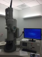
Transmission Electron Microscopy with the Philips CM 120
Location
- Center for Translational Research and Education (CTRE), room 173
Capable of collecting high quality, high resolution, 2-dimensional images of biological and non-biological specimens maintained at room temperature.
Instrument Features
- Tungsten electron gun (i.e. illumination source)
- Can be operated at 20, 40, 60, 80, 100, or 120 kV
- Equipped with twin objective lenses, a CompuStage specimen stage, and a single tilt sample holder
- Equipped with a BioSprint 16-megapixel digital camera
- Magnification range: 31× to 660,000×
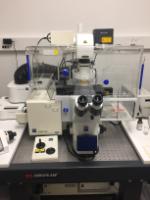
Multiphoton/Confocal Microscopy with the Leica TCS SP5
Location
Center for Translational Research and Education (CTRE), room 174
Capabilities
- Capable of collecting low and high magnification high quality, high resolution 2, 3, 4, and 5-dimensional (X, Y, Z, time, and spectrum) images
- Short-term live cell imaging experiments
- Semi-automated collection of 2- and 3-dimensional images from multiple X, Y, and Z locations over time
- Fluorescence recovery after photobleaching (FRAP), fluorescence resonance energy transfer (FRET), photo-activation (PA), photo-stimulation (PS), and photo-uncaging
- Tile stitching and image analysis
- Deep tissue (i.e., several hundred microns) imaging
Instrument Features
- Argon, HeNe, and titanium-sapphire (TiSa) pulsed (1-1.5 picoseconds duration) lasers
- Laser lines: 458, 476, 488, 496, 514, 543, 594, 633 nm
- Objectives: 10× dry, 20× dry, 40× oil immersion, 63× water immersion, 63× oil immersion
- Filter-free spectral detector system with 5 individually tunable channels
- Each detector is freely tunable over the entire visible spectrum
- The spectral range of the detector optics covers 350-900 nm with 5 nm spectral resolution and sub-micron spatial resolution
- The scanner can be run in conventional or resonant modes
- The conventional scanner can be scanned uni- or bidirectional with speeds ranging from 10-2800 Hz (2-3 fps at 512 × 512 pixels).
- The resonant scanner can be scanned uni- or bidirectional at 8000 Hz or 1600 Hz, respectively, with a maximum frame rate of 250 fps.
- Heated stage insert and controller
- Inverted microscope stand
- Digital zoom, 360° image rotation, and imaging multiple X, Y, and Z positions
- Equipped with a Z-galvanometer that allows for XZ scanning and fast XYZ scanning
- Scanning stage allows for stitching together images from large fields of view
- Time-lapse
- Leica Application Suite Advanced Fluorescence (LAS AF) software
- High confocal speed: 28 frames per second (fps) at 512 × 512 pixels
Image Analysis Workstation
Location
Center for Translational Research and Education (CTRE), room 175
Capabilities
- Interactive processing, visualization, and analysis of 3D and 4D microscopic images via Imaris software.
- State-of-the-art volume and surface rendering, object detection, tracking, and cell division tracking tools via Imaris software.
- Automated processing of image series in a batch.
- Automatic or semi-automatic segmentation, visualization, and quantification of filamentous structures via Imaris Filament Tracer module.
Instrument Features
- Dell Precision 7820 Tower base
- Windows 10 Pro for Workstation
- NVIDIA GeForce GTX 1080 graphic card
- Dual Intel Xeon gold 5122 3.6 GHz processors
- 128 GB (8×16GB) RAM
- Imaris 9.5 Basic Tracking with XT (XBT)
- Imaris Batch 9.5
- Imaris Filament Tracer 9.6
- Image J
Leica EM UC7 ultramicrotome
Location
Center for Translational Research and Education (CTRE), room 176
Capabilities
- Prepares consistent, high-quality semi-thin and ultrathin sections for transmission electron microscopy and light microscopy.
Instrument Features
- Thickness feed range from 1 nm to 15 µm.
- Cutting speed range from 0.05 to 100 mm/s.
- Eucentric movement allows for optimal knife approach to the sample.
- Outstanding LED illumination (top, back, and transmitted light; independent control of the illumination modes) yields outstanding visibility of the knife edge and block face.
- Fully motorized knife stage and auto-trim function.
- Vibration-decoupled gravity-stroke cutting arm for chatter-free sectioning.
- Touchscreen control unit.
Publications
2023
Talley S, Rademacher DJ, Campell EM. 2023. Inflammasome activation occurs in CD4+ and CD8+ T cells during graft-versus-host disease. Cell Death and Disease, 14(9):632. doi: 10.1038/s41419-023-06138-8
Ekyalongo RC, Flowers B, Sharma T, Zigrossi A, Zhang A, Quintanilla-Arteaga A, Singh K, Kastrati I. 2023. SELENOF controls proliferation and cell death in breast-derived immortalized and cancer cells, Cancers, 15(14):3671. doi: 10.3390/cancers15143671
Rademacher DJ. 2023. Potential for therapeutic-loaded exosomes to ameliorate the pathogenic effects of α-synuclein in Parkinson’s disease. Biomedicines, 11(4):1187. doi: 10.3390/biomedicines11041187
Wolfe AJ, Rademacher DJ, Mores CR, Evans RJ, Overholt T, Halverson T, Limeira R, Matthews C, Badlani G, Brubaker L, Walker SJ. 2023. Detection of bacteria in bladder mucosa of adult females. Journal of Urology, 209(5):937-949. doi: 10.1097/JU.0000000000003189
Choi JK, Naffouje SA, Goto M, Wang J, Cristov K, Rademacher DJ, Green A, Stecenko AA, Chakrabarty AM, Das Gupta TK, Yamada T. 2023. Cross-talk between cancer and Pseudomonas aeruginosa mediates tumor suppression. Communications Biology, 6(1):16. doi: 10.1038/s42003-022-04395-5
2022
Kim M, Nikouee A, Sun Y, Zhang QJ, Liu ZP, Zang QS. 2022. Evaluation of Parkin in the regulation of myocardial mitochondria-associated membranes and cardiomyopathy during endotoxemia. Frontiers in Cell and Developmental Biology, 10:796061. doi: 10.3389/fcell.2022.796061
Lehmann D, Sladek M, Khemmani M, Boone TJ, Rees, Driks A. 2022. Role of novel polysaccharide layers in assembly of the exosporium, the outermost protein layer of the Bacillus anthracis spore. Molecular Microbiology, 118(3):258-277. doi: 10.1111/mmi.14966
Yang F, Cabe M, Nowak HA, Langert KA. 2022. Chitosan/poly(lactic-co-glycolic)acid nanoparticle formulations with finely-tuned size distributions for enhanced mucoadhesion. Pharmaceutics, 14(1):95. doi: 10.3390/pharmaceutics14010095
Gupta Y, Sharma N, Singh S, Romero JG, Rajendran V, Mogire RM, Kashif M, Beach J, Jeske W, Poonam, Ogutu BR, Kanzok SM, Akala HM, Legac J, Rosenthal PJ, Rademacher DJ, Durvasula R, Singh AP, Rathi B, Kempaiah P. 2022. The multistage antimalarial compound Calxinin perturbates P. falciparum Ca2+ homeostasis by targeting a unique ion channel. Pharmaceutics, 14(7):1371. doi: 10.3390/pharmaceutics14071371
Burbidge K, Rademacher DJ, Mattick J, Zack S, Grillini A, Bousset L, Kwon O, Kubicki K, Simon A, Melki R, Campbell EM. 2022. LGALS3 (galectin 3) mediates an unconventional secretion of SNCA/α-synuclein in response to lysosomal membrane damage by the autophagic-lysosomal pathway in human midbrain dopamine neurons. Autophagy, 18(5):1020-1048. doi: 10.1080/15548627.2021.1967615
2021
Ghosh AK, Thapa R, Hariani HN, Volyanyuk M, Ogle SD, Orlaff KA, Ankireddy S, Lai K, Ziniauskaite A, Stubbs EB Jr, Kalesnykas G, Hakkarainen JJ, Langert KA, Kaja S. 2021. Poly(lactic-co-glycolic acid) nanoparticles encapsulating the prenylated flavonoid, Xanthohumol, protect corneal epithelial cells from dry eye disease-associated oxidative stress. Pharmaceutics, 13(9):1362. doi: 10.3390/pharmaceutics13091362
Yang F, Cabe MH, Ogle SD, Sanchez V, Langert KA. 2021. Optimization of critical parameters for coating of polymeric nanoparticles with plasma membrane vesicles by sonication. Scientific Reports, 11(1):23996. doi: 10.1038/s41598-021-03422-5
2020
Burbidge K, Zwikelmaier V, Cook B, Long MM, Balva B, Lonigro M, Ispas G, Rademacher DJ, Campbell EM. 2020. Cargo and cell-specific differences in extracellular vesicle populations identified by multiplexed immunofluorescent analysis. Journal of Extracellular Vesicles, 9(1):1789326. doi: 10.1080/20013078.2020.1786326
Gavini CK, Cook TM, Rademacher DJ, Mansuy-Aubert V. 2020. Hypothalamic C2-domain protein involved in MC4R trafficking and control of energy balance. Metabolism, 102:153990. doi: 10.1016/j.metabol.2019.153990
2019
Mastrodomenico V, Esin JJ, Graham ML, Tate PM, Hawkins GM, Sandler ZJ, Rademacher DJ, Kicmal TM, Dial CN, Mounce BC. 2019. Polyamine depletion inhibits bunyavirus infection via generation of noninfectious interfering virions. Journal of Virology, 93(14):e00530-19. doi: 10.1128/JVI.00530-19
Rademacher DJ, Cabe M, Bakowska JC. 2019. Fluorescent recovery after photobleaching of yellow fluorescent protein tagged p62 in aggresome-like induced structures. Journal of Visualized Experiments, 145: e59288. doi: 10.3791/59288
2018
Cabe M, Rademacher DJ, Karlsson AB, Cherukuri S, Bakowska JC. 2018. PB1 and UBA domains of p62 are essential for aggresome-like induced structure formation. Biochemical and Biophysical Research Communications, 503(4):2306-2311. Doi: 10.1016/j.bbrc.2018.06.153